
研究概要
我々の研究室では、MEMSや機械加工などのマイクロ・ナノ工学と超音波による機械力学を基盤技術として、ナノからマクロまでマルチスケールの“やわらかい”ロボットやデバイスを創出しています。これらを用いて、生体内への薬剤送達(Drug Delivery System)や組織工学/再生医療などの最先端医療に資する新たなプラットフォームを構築することを目指し、研究を展開していきます。
具体的には主に以下のトピックについて研究を実施しています。
- ヘルスケアと薬剤投与に資する生体内ハイドロゲルデバイスの創成
- 超音波照射デバイスによるナノ薬剤の低侵襲な生体投与
- ハイドロゲルを用いた3次元細胞組織の形成
- 超音波による細胞の非接触アクチュエーションシステムの開発
- ソフトロボティクスとAIの融合による細胞組織成熟システムの構築
我々の研究室では医学系や生物系の研究者と共に様々な医療や生命の課題に取り組み、これを機械工学の知識と技術により解決していきます。そして、上記以外のテーマ以外にも、生体や生物などに有効な機械工学の技術を用いた新たなプラットフォームについて、日々のディスカッションを通じて新たに研究を生み出していき,これまでにない革新的な技術やシステムを研究・開発します。新たなアイディアを形にしたい、マルチスケールで物作りがしたい、医療に役立つ研究がしたい、研究成果を海外で発表したいなど...世界をあっと言わせる研究を一緒に発信していきましょう!


ヘルスケアと薬剤投与に資する生体内ハイドロゲルデバイスの創成
ハイドロゲルは水を含む高分子ネットワークで構成され、その分子の特性により極めて多様な機能や性質を示します。特に、ハイドロゲルをマイクロ加工したハイドロゲルマイクロビーズは微小なサイズかつ比表面積が極めて大きいという特徴から、バルクでは得ることができない物質拡散の高速化や物質の均一性を有しています。こうした特徴から、薬剤送達(DDS)やバイオセンサなどの応用を見据えた研究が注目されています。特に、ハイドロゲルには周囲の環境に反応して性質を変化させることができることから、これらを組み合わせることで複数の機能を具備したインテリジェントデバイスを創成することが可能です。これを用いた生体内で作用する薬剤担体やインテリジェントロボットの構築を目指しています。
*この研究プロジェクトは,一部科研費の補助を受けて実施しました.
Simultaneous crosslinking induces macroscopically phase-separated microgel from a homogeneous mixture of multiple polymers
This paper reports a unique phase separation behavior, a simultaneous-crosslinking-driven phase sep- aration in co-gelation (SPSiC) core–shell microgel that spontaneously forms from a homogeneous pre- gel solution of multiple polymers. The SPSiC microgel, composed of an alginate shell and an N- isopropylacrylamide (NIPAM) core, were synthesized by a single fabrication step wherein a mixed pre-gel solution of sodium alginate and NIPAM monomer was ejected by centrifugation with photo- polymerization and ion crosslinking instantaneously. Phase separation was modeled by varying the degree of polymerization and the size of the polymer chain. Moreover, an implantable, multi-functional drug de- livery system combined with a transdermal glucose sensor was demonstrated with core–shell Janus SPSiC microgels. This work shows a macroscopic phase separation behavior, which occurs during the gelation process, and also provides a simple and unique methodology to create multifunctional bio-microprobes.

超音波照射デバイスによるナノ薬剤の生体投与
皮膚にある角質層は分子量500(約1〜2 nm)以上の物質の透過を妨げることで、我々のカラダを外敵から防御しています。しかしこれは同時に、ナノサイズの薬剤の経皮投与も妨げています。最近では、従来の低分子薬剤以外にも、特異性が高いことから安全性と薬効に優れた高分子薬剤が台頭してきました。高分子薬剤には、COVID-19で注目されている核酸医薬やがんの特効薬となる抗体医薬などがあり、これらを効率的に投与する方法の確立が期待されています。そこで、経皮薬剤投与のための超音波照射デバイスとハイドロゲルのナノカプセルを開発し、ニードルレスで安全に薬剤を投与する手法の構築を目指しています。本研究は薬剤投与以外にも、美容や遺伝子導入などにも応用が期待できます。
*この研究プロジェクトは,一部科研費および競輪の補助を受けて実施しました
Quantitative Analysis of Acoustic Pressure for Sonophoresis and Its Effect on Transdermal Penetration
Ultrasound facilitates the penetration of macromolecular compounds through the skin and offers a promising non-invasive technique for transdermal delivery. However, technical difficulties in quantifying ultrasound-related parameters have restricted further analysis of the sonophoresis mechanism. In this study, we devise a bolt-clamped Langevin transducer-based sonophoresis device that enables us to measure with a thin lead zirconate titanate (PZT) sensor. One-dimensional acoustic theory accounting for wave interaction at the skin interface indicates that the acoustic pressure and cavitation onset on the skin during sonophoresis are sensitive to the subcutaneous support, meaning that there is a strong need to perform the pressure measurement in an experimental environment replacing the human body. From a series of the experiments with our new device, the transdermal penetration of polystyrene, silica and gold nanoparticles is found to depend on the size and material of the particles, as well as the hardness of the subcutaneous support material. We speculate from the acoustic pressure measurement that the particles’ penetration results from the mechanical action of cavitation.

ハイドロゲルを用いた3次元細胞組織の形成
細胞は細胞同士や細胞周囲に接着することで細胞の塊を作り、これが集まることで組織を形成します。しかし、細胞だけを集積しても培養面に層状に積み重なるだけで、3次元組織を形成することはできません。そこで、ハイドロゲルを主材として細胞の足場を形成し、これを用いて細胞を組織化することで,マイクロサイズの細胞組織を形成することができる。この小さな組織を積み重ねてアセンブリすることで、巨大な立体組織を形成することが可能となる。その際に、複数種類の細胞を共培養することや血管網を設けることで、臓器に近い細胞組織を形成することができます。このような技術により形成した細胞組織は再生医療や創薬研究に用いるための細胞組織に応用することができます。
Microfiber-shaped building-block tissues with endothelial networks for constructing macroscopic tissue assembly
We describe a microfiber-shaped hepatic tissue for in vitro macroscopic tissue assembly, fabricated using a double coaxial microfluidic device and composed of cocultured Hep-G2 cells and human umbilical vein endothelial cells (HUVECs). The appropriate coculture conditions for Hep-G2 cells and HUVECs in the microfiber-shaped tissue were optimized by changing the thickness of the core and the cell ratio. The HUVEC networks were formed in the microfiber-shaped tissue following culture for 3 days. Using this microfiber-shaped tissue as a building block, two types of macroscopic assembled tissues were constructed—parallel and reeled tissues. In both tissue types, the connection of the HUVEC network across the adjacent microfiber-shaped tissues was established after 2days, because the calcium alginate shell of the microfiber-shaped tissue was enzymatically removed. Our approach could facilitate the generation of complex and heterogeneous macro- scopic tissues mimicking the major organs including the liver, kidney, and heart for the treatment of critically ill patients.

超音波による細胞の非接触アクチュエーション
超音波は生体内のセンシングや細胞・組織の破砕を非接触で行う方法として昔から使用されてきました。このような弱い/強い超音波を活用した技術以外にも、中間の出力を用いることで超音波をアクチュエータとして用いることで可能です。これにより、細胞を優しく非接触で操作できることから、細胞を汚染することなくマニピュレートすることができます。この超音波の特性を活かして細胞を損傷せずに剥離する手法を開発することや、細胞を組織化する手法を構築してきました。これにより、従来用いられてきた細胞操作技術を刷新する新たな技術を開発しています。
*この研究プロジェクトは,一部科研費の補助を受けて実施しました.
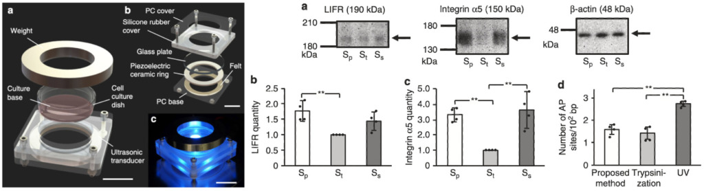
Enzyme-free release of adhered cells from standard culture dishes using intermittent ultrasonic traveling waves
Cell detachment is essential in culturing adherent cells. Trypsinization is the most popular detachment technique, even though it reduces viability due to the damage to the membrane and extracellular matrix. Avoiding such damage would improve cell culture efficiency. Here we propose an enzyme-free cell detachment method that employs the acoustic pressure, sloshing in serum-free medium from intermittent traveling wave. This method detaches 96.2% of the cells, and increases its transfer yield to 130% of conventional methods for 48 h, compared to the number of cells detached by trypsinization. We show the elimination of trypsinization reduces cell damage, improving the survival of the detached cells. Acoustic pressure applied to the cells and media sloshing from the intermittent traveling wave were identified as the most important factors leading to cell detachment. This proposed method will improve biopharmaceutical production by expediting the amplification of tissue-cultured cells through a more efficient transfer process.
Cell agglomeration in the wells of a 24-well plate using acoustic streaming
Cell agglomeration is essential both to the success of drug testing and to the development of tissue engineering. Here, a MHz-order acoustic wave is used to generate acoustic streaming in the wells of a 24-well plate to drive particle and cell agglomeration. Acoustic streaming is known to manipulate particles in microfluidic devices, and even provide concentration in sessile droplets, but concentration of particles or cells in individual wells has never been shown, principally due to the drag present along the periphery of the fluid in such a well. The agglomeration time for a range of particle sizes suggests that shear-induced migration plays an important role in the agglomeration process. Particles with a diameter of 45 mum agglomerated into a suspended pellet under exposure to 2.134 MHz acoustic waves at 1.5 W in 30 s. Additionally, BT-474 cells also agglomerated as adherent masses at the center bottom of the wells of tissue-culture treated 24-well plates. By switching to low cell binding 24-well plates, the BT-474 cells formed suspended agglomerations that appeared to be spheroids, fully fifteen times larger than any cell agglomerates without the acoustic streaming. In either case, the viability and proliferation of the cells were maintained despite acoustic irradiation and streaming. Intermittent excitation was effective in avoiding temperature excursions, consuming only 75 mW per well on average, presenting a convenient means to form fully three-dimensional cellular masses potentially useful for tissue, cancer, and drug research.

ソフトロボティクスとAIによる細胞組織成熟システムの構築
細胞組織は外側から機械的な刺激を付与することで筋肉を鍛えるように幼弱な細胞が成熟します。しかし、細胞は声を上げることができないことから、どのような刺激を付与することが細胞組織にとって最も適しているかを選定することは技術者の経験に委ねられています。このような状況を打開するために、AIを駆使して細胞に最も適した機械刺激を付与するための条件を探索する研究を行っています。
The paper is coming soon…